Showing posts with label birth injury. Show all posts

Tuesday, September 5, 2023

Expectant Mothers Face Limited or No Access to Prenatal Care

New Study Reveals: Millions of Expectant Mothers in the US Face Limited or No Access to Prenatal Care

A staggering 5.6 million women of a reproductive age in the United States are currently grappling with poor access to prenatal care, a concern exacerbated by the increasing rate of what's termed "maternity care deserts". According to a comprehensive research report by March of Dimes, 36% of counties across the US now fall under the classification of "maternity care deserts" (which means, they don’t have any birthing centers or obstetric service providers). Moreover, this recession and high cost of living are not helping things.

This report from March of Dimes has been published in the midst of a US healthcare sector that is wrestling with a significant lack of access to maternal health. Multiple independent studies have underscored that the US suffers from the most unfavorable maternal health outcomes and the most pronounced maternal health disparities among developed nations.

Dr. Elizabeth Cherot, President and CEO of March of Dimes, emphasized that a person's ability to have a healthy pregnancy and birth shouldn't be determined by their geographical location and access to consistent, high-quality care. With that said, this report illustrates that today these very factors jeopardize the lives of countless women in the US during pregnancy and childbirth.

Cherot further stated that their research demonstrates that maternity care is not adequately prioritized within the healthcare system, and immediate measures must be taken to ensure that all mothers receive the necessary care they deserve to facilitate healthy pregnancies and robust baby health.

The study noted an increased risks to the babies born in these areas. Necessarily, this will mean there is an increased risk of birth injuries such as cerebral palsy and HIE if proper prenatal and obstetrical care is not provided to the expectant mothers.


A Worrying Trend of Closure of Labor and Delivery Units

A striking 5.6 million women in their reproductive years live in counties where maternity care services are either absent or notably limited. This trend is largely attributed to the closure of labor and delivery units in hospitals. Since the initial coverage of this topic by March of Dimes in 2018, as many as 369 counties have witnessed the shutdown of their obstetric units and labor and delivery suites, constituting roughly one in ten counties.

An additional 70 counties in the US have now been branded as maternity care deserts following the closure of obstetric wards since the 2018 report. Collectively, more than one in three US counties now fall within the category of maternity care deserts, characterized by the absence of hospitals or birthing centers providing obstetric care, and devoid of obstetric service providers.

The authors of the report attribute the closure of obstetric wards to factors such as escalating operational costs. The report cites statistics from the American Hospital Association to highlight that only about 50% of births in maternity care deserts are reimbursed through Medicaid, which offers lower compensation compared to private insurance.

This renders obstetric wards prime targets for closure in hospitals dealing with narrow profit margins.

Dangers to the Health of the Expectant Mother and the Baby

Regrettably, this national problem is severely detrimental to the health of the expectant mother. Researchers underline that over 32 million women in their reproductive years are susceptible to compromised health outcomes due to their inability to access reproductive healthcare services. This issue is most acute in rural states.

The presence of chronic illnesses substantially contributes to health outcomes, the study authors suggest. Eight out of ten maternity care deserts have a significant concentration of expectant mothers struggling with chronic ailments, some of which exert a direct impact on birthing conditions like preeclampsia (maternal high blood pressure) and premature birth. Many of these preventable conditions can lead to a birth injury like cerebral palsy or hypoxic ischemic encephalopathy (HIE).

Social determinants of health and chronic illnesses are even more prevalent in regions that are already marked as maternity deserts, which, in turn, place an added burden on expectant mothers who have limited access to healthcare options.


The Underlying Causes of Maternity Deserts

The states registering the highest incidence of maternity care deserts include North Dakota, South Dakota, Alaska, Oklahoma, and Nebraska. These states predominantly feature rural populations, as revealed in the March of Dimes report.

For instance, in North Dakota, a substantial 71.3% of rural residents are situated at least 30 minutes away from a maternity care hospital, with an average travel distance of 54.7 miles, Dr. Cherot stated. In terms of care quality, New Mexico displays the highest rate of substandard prenatal care, closely trailed by Hawaii, which is a state that’s expensive to live in.

It’s noteworthy that a socio-economic and racial dimension is also at play. Dr. Cherot pointed out that Black and Native American women experience the most unfavorable outcomes, coupled with the highest incidence of inadequate prenatal care.


Ignoring Prenatal Care Due to Restricted Choices

An expectant mother residing in a maternity desert is compelled to visit the nearest county where care is accessible. In a few cases, this might involve crossing just a nearby county boundary. For many others, this could entail hours of travel, a situation fraught with danger during active labor or a medical emergency. It is well-documented that a leading cause of HIE and cerebral palsy is a delay of delivery when the baby is suffering from fetal distress. Forcing an expectant mother to travel long distance to deliver her child only increases the risks of these serious birth injuries.


Expectant mothers in these maternity deserts often attend a fraction of prenatal appointments.

Some expectant mothers may consult community midwives who lack formal licensing and consistent training. Alternatively, they might head to the nearest emergency room whenever they sense something is amiss, without the benefit of continuous care from a regular provider.

Even more alarmingly, a number of women may bypass prenatal care altogether. Dr. Cherot remarked that expectant mothers who forego prenatal care face three times higher odds of experiencing adverse outcomes.

In some instances, women go into labor while en route to a hospital. Dr. Kristy Acosta, a Family Medicine/OB practitioner at the Brownfield Regional Medical Center in Texas, reported that it's not uncommon for her to receive a call that a mother has given birth at a gas station. She then awaits their arrival at the emergency room.

According to the article, Kali Bautista, a resident of Terry County, Texas, delivered her child while dwelling in a maternity desert within the state. Her hometown is situated 30 to 45 minutes away from Lubbock. She shared her concerns saying that one of her chief fears was what would happen if she went into labor during the journey.

Staffing Shortages and Obstetrician Burnouts

The closure of hospitals or maternity units can be attributed to a variety of factors, predominantly revolving around a shortage of trained staff. According to March of Dimes' press release, obstetricians exhibit one of the highest burnout rates within the healthcare industry, complicating the process of attracting and retaining staff.

In just one year, the count of birthing hospitals has dwindled by 4%. Since 2018, as many as 301 birthing units have ceased operations. Experts indicate a serious shortage of qualified OB-GYNs across the United States. Moreover, as a significant proportion of doctors are nearing their retirement age, a wave of retirements is imminent in the upcoming years. And there is a clear decline in the number of new OB-GYNs entering the workforce to fill the void.

While midwives offer valuable support in the birthing process, they do not engage in surgical procedures like operative vaginal deliveries, cesarean sections, cervical cerclage (suturing the incompetent cervix to prevent premature birth), handling the birth of a breech baby, or other complicated aspects of obstetric care.


Escalating Costs of Care and Inadequate Financial Reimbursement

Increasing costs of prenatal care and insufficient reimbursement also contribute to this problem, as shown by the March of Dimes report. Only about half of the births in maternity care deserts are covered by Medicaid, as per the American Hospital Association's data. This implies that hospitals receive lower reimbursement rates, leading to financial losses when offering obstetric care.

Dr. Cherot stated that the issue ultimately boils down to economics. Purely for economic reasons, the healthcare system in the country is failing to accord the necessary priority to the well-being of mothers and babies. Maternal health is a concern that reverberates throughout the society, and the March of Dimes report underscores that each one of us has a role to play in safeguarding the health of both expectant mothers and babies.


Miller Weisbrod Olesky has a long history of helping victims of cerebral palsy and HIE

Whether the birth injury is caused by a lack of proper prenatal care, a delay due to the lack of proper transfer from a ill-equipped hospital or the mistakes of the medical team during labor and delivery or shortly after childbirth, our experienced cerebral palsy and HIE birth injury attorneys have helped families all across the United States that are struggling with the reality and costs of raising a disabled child.

Families have the right to know if their child’s cerebral palsy or HIE were caused by mistakes of doctors, nurses and hospitals. For absolutely no initial cost to you and your family, we will conduct a detailed review of the medical records and all other available information to determine if your child’s birth injury was avoidable. There were never be a fee unless and unti we receive a successful money settlement for your child.

Tuesday, June 14, 2022

Dallas Birth Injury Lawyers

Dallas Birth Injury Lawyers Fighting For Your Baby's Future

Our national birth injury law firm helps families who face the devastating consequences of birth injuries caused by medical malpractice and negligence.

The Dallas Birth Injury Lawyers of Miller Weisbrod Olesky focus on providing experienced, skilled representation to get the best results for our clients. Our track record of successful settlements and verdicts speaks for itself.

Birth Injuries Strike the Most Innocent of Victims

Expectant mothers and their babies rely on medical professionals to give them the care they need. Birth injuries can happen when those doctors, nurses, midwives, and hospitals fail.

Dallas Birth Injury Lawyers

Before a baby is born, it’s crucial for doctors to identify and treat any conditions that arise like:

  • Serious maternal medical conditions, like diabetes, high blood pressure, and infections
  • Signs the baby is not growing as expected
  • Abnormal heartbeats
  • Decreased fetal activity
  • Abnormal fetal presentation (breech)
  • Risk of premature delivery
Fetal Heart Rate Monitoring Lawyers

During labor and delivery, situations arise that could harm the baby if medical professionals quickly provide the proper care:

Dallas Fetal Distress Lawyers

Finally, babies can suffer birth injuries after they are born when doctors and nurses don’t treat the following conditions:


Was your child injured at birth? If so, our Dallas Birth Injury Lawyers can discuss your situation during a free consultation. Just call (888) 987-0005 to set up your free consultation.

Birth Injuries Take a Toll on Children and Their Families

Dallas Cerebral Palsy Lawyers

Depending on how badly your child was injured, you might be facing years of medical treatment and therapy. Many children with birth injuries need the following kinds of care:


Dealing with a birth injury is emotionally and financially exhausting. Registered Nurses and Nurse-Attorneys Are a Vital Part of Our Birth Injury Team … and Yours

Miller Weisbrod Olesky's Nurse Attorneys

As Dallas Birth Injury & Cerebral Palsy Attorneys, we fight to get justice for our clients. We believe in holding negligent medical professionals accountable for their negligence. We do this by:

  • Discussing your case with you in detail
  • Using our extensive resources to investigate your baby’s records thoroughly
  • Providing nurse-attorneys and nursing staff who understand what has happened medically
  • Consulting with experts who understand how you and your child should have been treated
  • Seeking compensation for your baby’s injuries from the people who caused them

The compensation our clients receive in birth injury lawsuits makes a difference. They can better care for their child while also covering other expenses like childcare and lost wages.

Miller Weisbrod Olesky Birth Injury Lawyers

Free Consultations

We do not collect a fee until we win your case.

You’re facing some challenging decisions – whether someone caused your baby’s birth injury and how to hold them accountable. As experienced Dallas Birth Injury Attorneys, we have obtained verdicts and settlements for clients like you. The millions of dollars our clients received were used for therapies, treatments, assistive devices, caregivers, and planning for the future. Don't hesitate to get in touch with us today to discuss your unique circumstances.

Wednesday, March 16, 2022

Learning About Cerebral Palsy

cerebral palsy home health

Cerebral Palsy is a congenital disorder of movement, muscle tone, or posture. Cerebral palsy is due to abnormal brain development, often before birth. CP is the most common motor disability in childhood.

Cerebral means having to do with the brain. Palsy means weakness or problems with using the muscles. Cerebral palsy is a group of physical disorders that permanently affect:

  • Posture
  • Balance
  • Movement
  • Ability to control movement
  • Communication
  • Eating
  • Sleeping
  • Learning

What Causes Cerebral Palsy?

The causes of cerebral palsy can be more difficult to grasp if medical errors are involved. Cerebral palsy can be caused by doctors or nurses failing to respond to fetal distress, failing to perform a timely C-section or other errors during the labor and delivery process or care shortly after birth of a child. These errors can constitute medical malpractice.


Symptoms Of Cerebral Palsy

The symptoms of cerebral palsy are primarily physical. However, injuries to an infant’s brain, including hypoxic-ischemic encephalopathy (HIE), are the primary cause of CP. Cerebral actually means brain, while palsy means muscle weakness.Symptoms vary widely among children with cerebral palsy. Some signs may show up soon after delivery, while milder cases may not be diagnosed until later, usually by two to five years of age.

The two general classifications of symptoms are:

  • Decreased muscle tone (Hypotonia) – Infants with hypotonia may seem floppy. Brain damage can cause hypotonia
  • Increased muscle tone (Hypertonia) – Muscles become stiff and challenging to move. Hypertonia also can be caused by brain damage
hypertonia and hypotonia

Between ages 6 months and 12 months, an infant with cerebral palsy might exhibit the following symptoms:

  • Missing physical development milestones
  • Unable to roll over
  • Difficulty moving hands
  • Keeping one hand fisted while reaching with the other
  • Lopsided crawling
  • Not crawling on all fours

Adults with cerebral palsy might exhibit additional symptoms, including:

  • Lack of muscle coordination
  • Difficult performing precise movements
  • Difficulty walking
  • Stiff muscles
  • Exaggerated reflexes
  • Shaking, twitching, or other involuntary movements
  • Drooling or having trouble swallowing
  • Difficulty speaking

Types Of Cerebral Palsy

Cerebral palsy is a complex disorder where brain damage causes physical disabilities. The wide range of symptoms and degrees of severity complicate diagnosis and treatment, as well as perception.

types of cerebral palsy

Within the cerebral palsy diagnosis, there are four basic types of CP:

  • Spastic cerebral palsy
  • Ataxic cerebral palsy
  • Dyskinetic cerebral palsy
  • Mixed cerebral palsy

Spastic cerebral palsy is the most common type of cerebral palsy. In fact, roughly 80% of people with CP are diagnosed with a form of spastic CP. People with spastic cerebral palsy exhibit uncontrollable, involuntary limb movements and tight muscles that inhibit movements, causing bone and joint deformities.

  • Diplegia or Diparesis: This type of CP indicates that an individual has severe muscle stiffness, particularly in their legs. Their arms might be affected very little or not at all.
  • Hemiplegia or Hemiparesis: Here, disability affects only one side of an individual’s body. Typically, this type of CP affects a person’s arms more than their legs.
  • Quadriplegia or Quadriparesis: This is the most severe form of spastic cerebral palsy because it affects a person’s arms, legs, trunk, and face. Also, people with this form of CP usually have intellectual disabilities and suffer from seizures and speech, hearing, and vision problems.

Dyskinetic cerebral palsy is the second most common form of cerebral palsy. Research indicates dyskinetic cerebral palsy is caused by “non-progressive lesions to the basal ganglia or thalamus or both.” Frequently, birth trauma due to medical errors during labor and delivery or immediately following birth (called “neonatal negligence”) causes these injuries.

Since dyskinetic cerebral palsy may affect the face and tongue, it may be difficult to talk, suck, and swallow. However, the muscle tone of children with dyskinetic CP often varies from day to day and even from hour to hour. Doctors recognize three forms of dyskinetic CP based on an individual’s symptoms:

  • Dystonic: Dystonic symptoms include twisting, repetitive movements that are more prominent when a child is tired or emotional. For some children, pain levels increase with increased movement. These symptoms affect activities of daily life, quality of life, and participation in social activities.
  • Athetoid: Characterized by slow, writhing movements, athetoid’s primary symptoms include abnormal posture, impaired coordination, and minimal control over movement. A child with athetoid symptoms might be unable to maintain posture. Other limitations include trouble with standing, sitting, and hand movements. Athetoid dyskinetic cerebral palsy can be caused by bilirubin encephalopathy or hypoxic-ischemic encephalopathy (HIE).
  • Choreoathetosis: Someone with chorea symptoms will have movements that are clumsy, abrupt, and unpredictable. Wild, violent movements are possible with more severe forms of choreoathetosis cerebral palsy. Chorea can interfere with movements, speech, and swallowing.

Dystonia and choreoathetosis are often found together.

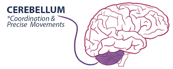
Ataxic Cerebral Palsy refers to issues with balance and coordination. Children with this form of CP might appear unsteady and shaky. Their movements might seem disorganized and jerky to other people.

It’s believed that ataxic cerebral palsy is the result of damage to the cerebellum. This part of the brain is responsible for controlling movement and plays a part in “cognition, emotional processing, and social behavior.”

cerebellum

Mixed cerebral palsy is the diagnosis when children have a combination of cerebral palsy symptoms. Typically, this means that brain injury has occurred in several parts of the brain. Currently, spastic-dyskinetic CP is the most common type of mixed cerebral palsy.


Cerebral Palsy Treatments

There’s no cure for cerebral palsy. But that doesn’t mean there’s no way to alleviate symptoms and improve abilities. In fact, treatments and therapies that are currently available can help someone with cerebral palsy live as fully as possible.

Typical goals for a child with CP include:

  • Preventing or reducing physical symptoms
  • Preventing or reducing physical symptoms
  • Improving the child’s mobility
  • Optimizing the child’s overall health
  • Maximizing success and independence

Treatment plans for people with cerebral palsy may include medications, surgeries, and therapies. Also, medical equipment and assistive technology often play an essential part in the daily life of a child with CP.


Cerebral Palsy Medications

Medical professionals might administer medications orally, in IVs, or through muscle injections. The drugs prescribed for people with cerebral palsy fall into the following categories:

  • Anti-spasmodic: These drugs help reduce muscle spasms, spasticity, and tight muscles. Examples: Baclofen, benzodiazepines like diazepam, lorazepam. Dantrolene sodium (Dantrium and Revonto). Diazepam (Valium). Tizanidine or Flexeril (muscle spasms). Botulinum toxin (injected). Intrathecal baclofen (implantable pump)
  • Anticonvulsant: Medical professionals sometimes prescribe anticonvulsants for children who experience seizures. Examples of anticonvulsants medications include Clonazepam/Klonopin (benzodiazepines), Dilantin (phenytoin), Tegretol (carba-mazepine)
  • Anticholinergic: Doctors sometimes prescribe anticholinergics to patients with involuntary muscle movements and excessive drooling. Examples are benztropine mesylate, Robinul, and Sinemet
  • Anti-inflammatory: These medications might be administered for pain relief. Reducing inflammation also helps control the chronic pain that people with CP often face. Examples: Nonsteroidal anti-inflammatory drugs, steroids

Doctors also often prescribe anti-depressants for people suffering from depression. In addition, recent studies indicate that medications containing cannabidiol (CBD) may have a positive effect on children with cerebral palsy. However, research is ongoing.


Cerebral Palsy Surgeries

Sometimes surgery is the best option to treat a child’s cerebral palsy symptoms, particularly those associated with motor skill deficits.

  • Selective dorsal rhizotomy: Pediatric orthopedic surgeons use this surgery to “improve communication between the spine and muscle.” This surgery can help decrease muscle tone and stiffness problems
  • Intrathecal baclofen pump: Baclofen is used to treat muscle spasms, which is a common symptom of cerebral palsy. Some patients benefit from having an intrathecal baclofen pump surgically inserted into their abdominal wall. The pump then sends baclofen directly to the patient’s spine
  • Orthopedic surgeries: Some children and adults with cerebral palsy benefit from surgery used to ease tight muscles and correct problems with bones and joints
  • Surgeries to Address Other Conditions: Cochlear implants can improve or restore a patient’s hearing. Many people with cerebral palsy suffer from gastrointestinal problems that can be eased with surgery. Finally, nasogastric tubes and gastronomy tubes help when feeding issues arise

Cerebral Palsy Therapies

Therapies like the following can help people with cerebral palsy improve and retain capabilities.

assisted devices and equipment

  • Physical Therapy: Typically, children learn how to sit, walk, and use orthotics with the help of a physical therapist. Other benefits include stretching muscles, improving mobility, and preventing or reducing muscle contractures
  • Occupational Therapy: This type of therapy teaches children how to handle activities of daily living. For example, children may learn how to get dressed, bathe, and eat. Therapists help the children make the most of their abilities, which can improve the child’s self-confidence. Occupational therapists tend to focus more on fine motor skills and hand-eye coordination
  • Speech and Language Therapy: Many people with cerebral palsy experience speech and communication issues. This type of therapy helps people improve their ability to speak and addresses muscle control problems with the tongue and mouth. Some children learn new ways to communicate, like sign language or computer software
  • Recreational Therapy: Engaging in recreational activities can help a child with cerebral palsy improve their physical and intellectual skills. Also, parents often see improvement in self-esteem and emotional well-being
  • Alternative Therapies: Children may also benefit from other, complementary forms of therapy, including:

    • Hippotherapy: Riding and interacting with specially-trained horses sometimes improves a child’s balance, especially children with pelvic bone disorders. This therapy also helps with other activities involving balance and postural alignment, including walking, jumping, and using stairs
    • Hyperbaric oxygen therapy (HBOT): This therapy exposes children to pressurized air or oxygen while in a special chamber. With higher air pressure, lungs can take in extra oxygen that is then used throughout the body to promote healing. HBOT is often used for people with traumatic brain injuries.

The parents of children with cerebral palsy often want answers. They want to know what happened to harm their child’s brain. Was it preventable?

Birth Injury Cerebral Palsy Attorneys

Our dedicated birth injury lawyers want to help you find those answers. We diligently investigate the facts and hold responsible parties accountable by pursuing medical malpractice claims against them. The compensation our clients receive helps them pay for their child’s current and future medical treatment, assistive technology and equipment, and the other expenses associated with caring for a child with cerebral palsy.

At Miller Weisbrod, a team of committed professionals use our detailed case review process to assess your potential claim. They start by learning more about you and your child. Then we gather medical records to determine what happened before, during, and after your delivery. We call in skilled medical experts who review your records and let us know if they think medical errors could have caused your child’s injuries.

The medical review of your case and the consultation are free. We only receive payment when you do when, and only when, a money settlement is reached for your child.

If your family has been affected by a birth injury such as cerebral palsy caused by a doctor's mistake or delivery room negligence, we invite you to contact our firm to discuss your case in a free initial consultation.

Our birth injury attorneys have the proven record, experience, and resources to help you achieve the compensation you deserve. Contact our offices today at (888) 987-0005 for your initial free consultation. You may also complete the form on this page to contact our support team.

Tuesday, September 14, 2021

Newborn Resuscitation

Dallas Birth Injury Lawyers
During pregnancy the baby is dependent on the umbilical cord and placenta for oxygen delivery. 10% of newborns require some form of assistance to start breathing on their own and 1% requires extensive medical intervention known as neonatal resuscitation.

Neonatal resuscitation refers to emergency intervention techniques employed immediately after childbirth to assist babies who are not able to breathe independently after birth. If a newborn cannot begin breathing independently right away they are at risk of birth asphyxia which can cause serious brain injury and even death.

When abnormal breathing is observed after delivery, doctors (neonatologists/pediatricians) and neonatal nurses will have a very short window of time to intervene to avoid harm to the baby. Anticipating the potential need for neonatal resuscitation is often critical. The leading risk factor for newborn respiratory problems is prematurity. Another common cause of the need for neonatal resuscitation is birth injury from inadequate oxygen to the unborn baby during the labor and delivery process.

Risk factors and Symptoms of Neonatal Resuscitation:
  • Maternal hypertension or cardiovascular disease
  • Multiples (twins)
  • Maternal drug/alcohol usage
  • Trauma during birth
  • Maternal age over 40
  • Fetal macrosomia
  • Meconium stained amniotic fluid
  • Maternal infection
  • Placental abruption
Neonatal Guidelines
Anticipation and Preparation: Newborn resuscitation depends heavily on proper planning and readiness of the team. Effective team behaviors, such as anticipation, communication, briefing, equipment checks, and assignment of roles, result in improved team performance and neonatal outcome.

The obstetrician or maternal fetal medicine doctors and the labor and delivery nurses are required to arrange a team to be present if there are signs that neonatal breathing assistance or neonatal resuscitation may be required. The hospital should then respond by sending a team to the delivery room.

This team may consist of neonatal nurses and/or doctors that will be either a neonatologist or pediatrician, depending upon the size and sophistication of the hospital. If a neonatal team is not requested, or is not assembled prior to delivery, then valuable minutes may be lost while the baby is not breathing.

Even if a team is not present at the time of delivery, the labor and delivery nurses and obstetrician delivering the baby must urgently call a team if a baby is unexpectantly born without breathing or not breathing properly. Every minute counts when a baby is not breathing!

Positive-pressure ventilation and Oxygen therapy (PPV): PPV remains the primary method for providing support for newborns that are apneic, bradycardic, or demonstrate inadequate respiratory effort. Pulse oximetry is used to guide oxygen therapy and meet oxygen saturation goals. This can include the intubation of the baby. A timely intubation will save a baby that is not breathing from any further damage. With proper oxygen therapy, a rise in heart rate is the most important indicator of effective ventilation and response to resuscitative interventions.

Chest compressions: If the heart rate remains less than 60/min despite 30 seconds of adequate PPV, chest compressions should be provided. The suggested ratio is 3 chest compressions synchronized to 1 inflation (with 30 inflations per minute and 90 compressions per minute) using the 2 thumb–encircling hands technique for chest compressions.

Vascular access: When vascular access is required in the newly born, the umbilical venous route is preferred. When intravenous access is not feasible, the intraosseous route may be considered.

Medications: If the response to chest compressions is poor, it may be reasonable to provide epinephrine, preferably via the intravenous route. This can help restart a baby’s heart and must be done timely!

Volume expansion: Failure to respond to epinephrine in a newborn with history or examination consistent with blood loss may require volume expansion.

Knowing the limitations: Doctors and nurses who provide neonatal resuscitation are faced with many challenges with respect to the knowledge, skills, and behaviors needed to perform effectively. These medical providers must be adequately trained on neonatal resuscitation methods and interventions. Neonatal resuscitation will always benefit from ongoing booster training, briefing, and debriefing.

Slow or improper performance of these procedures, and failure to perform procedures when indicated, can critically deprive adequate oxygen and blood flow to the baby. There are very specific guidelines published for medical professionals regarding what should be done in situations requiring neonatal resuscitation. Failure to follow acceptable standards and guidelines relating to neonatal resuscitation is medical negligence or medical malpractice.

Deprivation of oxygen and blood can lead to disability and permanent brain injuries, including hypoxic-ischemic encephalopathy (HIE), cerebral palsy, periventricular leukomalacia (PVL), and brain bleeds. The type and severity of the brain damage depend on many factors, including how long the baby was deprived of adequate blood flow and oxygen.

The Texas Birth Injury Attorneys at Miller Weisbrod understand the significant impact a birth injury will have not only on your child's life, but also your own. As experienced Birth Injury Lawyers, we fight hard for answers and justice if a delivery room error or doctor's negligence caused your child's birth injury.

If your child suffered a serious injury or lifetime disability due to a mistake before, during or after delivery, a lawsuit may be the best way to find out once and for all what happened and who should be held responsible. Contact us today to schedule a free consultation with an experienced birth injury lawyer. We encourage you to call our offices today at 214.987.0005 or toll free at 888.987.0005. You may also contact us by filling out the form on this page for answers to your important questions or to schedule an appointment.

Thursday, September 9, 2021

What Is Neonatal Hypoglycemia

Neonatal Hypoglycemia (NH) is one of the most common metabolic problems in babies, and severe NH is one of the leading causes of a brain injury to your baby. When your baby is born, the doctors will often order your baby’s blood glucose (sugar) to be monitored even if your baby is completely healthy. This is because of danger that hypoglycemia can pose even to healthy babies.

When your baby’s blood does not deliver enough glucose (sugar) to the brain, the lack of sugar can lead to serious consequences to your baby’s young brain because an infant’s developing brain tissue depends on a steady supply of glucose as its main source of fuel for making the transition to life. When a baby’s brain doesn’t receive a sufficient amount of glucose, the cells in the brain begin to die, and this can lead to permanent brain damage, developmental delays, and long-term loss of brain tissue.

Known Conditions That Cause NH:
  • Babies who are unusually small or large for gestational age
  • Babies who were born to diabetic mothers
  • Babies who have a history of asphyxia (HIE) or stress in the womb
  • Babies with low thyroid hormone levels (hypothyroidism)
  • Babies who have certain rare genetic disorders
  • Preterm babies: may have poor nutrient reserves and immature hormone systems that can increase their risk

Long-Term Effects of neonatal hypoglycemia:
If neonatal hypoglycemia goes undiagnosed and/or untreated for too long, there is a chance for long-term injury:
  • Brain damage
  • Cerebral palsy
  • Learning disabilities
  • Developmental disabilities
  • Epilepsy/seizures
  • Vision problems
  • Neuropsychiatry disorders

There are two types of neonatal hypoglycemia, transient (short-term) and persistent (long-term). Babies who have transient NH typically have a deficiency of glycogen stores at birth. This is common in babies that are born premature, who are small for gestational age, or experienced birth asphyxia.

Some infants with low blood sugar may not show symptoms. Routine blood tests are done following birth to check blood sugar levels. If signs and symptoms do surface:
  • Bluish-colored skin (cyanosis) or pale skin
  • Breathing problems, such as rapid breathing (tachypnea), pauses in breathing (apnea), or a grunting sound
  • Irritability or listlessness
  • Loose or floppy muscles (hypotonia)
  • Vomiting or poor feeding
  • Weak or high pitched cry
  • Tremors, shakiness, sweating, or seizures

Treating Neonatal Hypoglycemia (NH)
There are several treatment options available for the management of neonatal hypoglycemia; however, selecting the appropriate intervention can be challenging as the underlying cause may take weeks to diagnose. Therefore, during the diagnostic process, it is important to prevent or minimize periods of hypoglycemia in an effort to mitigate potential adverse neurological outcomes caused by insufficient glucose availability for optimal brain function.
Treatment includes giving the baby a fast-acting source of glucose. This may be as simple as a glucose and water mixture or formula as an early feeding. Or your baby may need glucose given through an IV. The baby's blood glucose levels are checked after treatment to see if the hypoglycemia occurs again.

Aggressive management of neonatal hypoglycemia is important as impaired neurodevelopmental outcomes are recognized in this patient population.

Contact our Birth Injury Attorneys
The Texas Birth Injury Attorneys at Miller Weisbrod understand the significant impact a birth injury will have not only on your child's life, but also your own. As experienced Birth Injury Lawyers, we fight hard for answers and justice if a delivery room error or doctor's negligence caused your child's birth injury.

To discuss your case with an experienced Birth Injury Lawyer, please fill out the contact form on this page to schedule an appointment. You can also call us directly at 214-987-0005 or toll free at 888-987-0005.

Tuesday, September 7, 2021

Signs and Symptoms of Fetal Distress

Fetal Distress
Fetal distress refers to signs before and during childbirth indicating that the fetus is not well. Fetus is the medical term for a baby prior to delivery. Fetal distress is a complication of labor. It occurs when the baby has not been receiving enough oxygen (birth asphyxia).

The baby reacts at the onset of asphyxia with a series of responses, a regulated redistribution of blood flow that serves to limit the effects of oxygen limitation in vital organs. This enables the baby to survive asphyxia intact unless the insult is profound or prolonged. Each of these stresses produces characteristic fetal heart rate patterns: late decelerations, variable decelerations, or prolonged bradycardia.

Common Conditions
The main sign doctors and nurses look for to determine fetal distress is a non-reassuring heart rate on the fetal monitoring strips. There are a number of conditions that can occur during labor and delivery that may lead to the fetal heart rate to become non-reassuring.

  • Anemia (most prevalent obstetric condition seen behind non-reassuring fetal status)
  • Oligohydramnios (a condition in which there is a lower level of amniotic fluid around the baby)
  • Pregnancy Induced Hypertension (PIH)
  • Post-term pregnancies (42 weeks or more)
  • Intrauterine Growth Restriction (IUGR)
  • Meconium-stained amniotic fluid (a condition in which meconium, a baby’s first stool, is present in the amniotic fluid which can block baby’s airways)

Symptoms of Fetal Distress
Common Signs of Fetal Distress
Decreased Fetal Movement in the Womb
Movement within the womb is an important indicator of the baby’s health. Some regular pauses in movement are normal because babies sleep in the womb. If the baby becomes less active or ceases to move, this may be a cause for concern.

Abnormal Fetal Heart Rate
To observe an unborn baby’s heart rate, labor and delivery nurses and obstetricians can use either an external or internal fetal monitoring device. External monitoring is done through a belt-like device that can be strapped around a mother’s abdomen, while internal monitoring involves attaching an electrode to the baby’s scalp. Internal monitoring provides the most accurate monitoring.

During the actual labor process, the important document the nurses and doctors must watch are fetal monitoring strips (these are the print outs from the monitor). These strips will have some of the earliest and most telling signs of the potential for fetal distress. Nurses and doctors during the labor and delivery process look for certain heart rate patterns that are recognized signs of fetal distress.
Some fetal heart rate patterns are known indicators of fetal distress. These are called non-reassuring heart rate patterns. The following fetal heart rate patterns are examples of nonreassuring patterns and warrant further investigation and medical intervention:

  • An abnormally fast heart rate (tachycardia)
  • An abnormally slow heart rate (bradycardia)
  • Abrupt decreases in heart rate (variable decelerations)
  • Late returns to the baseline heart rate after a contraction (late decelerations)

Abnormal Amniotic Fluid Level
The amount of amniotic fluid can be determined using a variety of ultrasound methods, including a qualitative assessment, the single deepest pocket (SDP), and the amniotic fluid index (AFI). The qualitative assessment is fairly subjective. The ultrasonographer scans the uterus and reports whether the amniotic fluid volume appears to be low, normal, or high, based on their own experience.

If there is abnormally low amniotic fluid, this is a condition called oligohydramnios, which can lead to oxygen deprivation and birth injuries like Hypoxic Ischemic Encephalopathy (HIE) and cerebral palsy (CP).

Abnormal Results of Biophysical Profile (BPP)
A baby’s biophysical profile (BPP) is also often taken if the results of a Nonstress Test (NST) are nonreassuring. In addition to the NST results, the BPP includes an ultrasound to assess fetal movement, breathing, tone, and amniotic fluid volume.

Vaginal Bleeding
Vaginal bleeding is common during pregnancy. Bleeding can also be an indication that something is wrong with the pregnancy. One particularly dangerous example is placental abruption, which occurs when the placenta tears away from the womb. This causes the baby to be deprived of oxygen.

A placental abruption and other placental problems that cause bleeding require very close monitoring, and in many cases, the mother should be admitted to the hospital and given an emergency C-section.

Cramping
Cramping is normal during pregnancy. As the baby grows, the uterus needs to expand. In some cases cramping is an indication of something more serious, such as miscarriage, placental abruption, preeclampsia, a urinary tract infection, or preterm labor.

Maternal High Blood Pressure (Preeclampsia)
Preeclampsia happens when a woman who previously had normal blood pressure suddenly develops high blood pressure* and protein in her urine or other problems after 20 weeks of pregnancy. Women who have chronic hypertension can also get preeclampsia.

Insufficient or Excessive Maternal Weight Gain
Experts believe that for women with a healthy pre-pregnancy weight, a weight gain of anywhere between 25 and 35 pounds is normal during pregnancy.

Fetal Distress that is not recognized and responded to is negligence and this can cause a birth injury including Cerebral Palsy and other brain injuries to a baby.

Healthcare providers, including both labor/delivery nurses and doctors, must recognize and react to signs of fetal distress. This may include supplemental oxygen, turning the mom and signs of the distress that do not improve or indicate profound fetal distress they must deliver the baby quickly including performing a Cesarean Section (c-section).

There is nothing more traumatic than your child suffering a birth injury. Many birth injuries cause serious medical problems that could end in lifetime disability. The Texas Birth Injury Attorneys at Miller Weisbrod understand the significant impact a birth injury will have not only on your child's life, but also your own. As experienced Birth Injury Lawyers, we fight hard for answers and justice if a delivery room error or doctor's negligence caused your child's birth injury.

If your child suffered a serious birth injury or lifetime disability due to a mistake before, during or after delivery, a lawsuit may be the best way to find out once and for all what happened and who is to blame. Contact our Birth Injury Lawyers today to schedule a free consultation. Call Miller Weisbrod, Attorneys At Law today at 214.987.0005 or toll free at 888.987.0005 for a free consultation.

Wednesday, August 11, 2021

Neonatal Therapeutic Hypothermia

Neonatal Therapeutic Hypothermia (Cooling)

Treatment for Hypoxic-Ischemic Encephalopathy (HIE)

What is Hypoxic-Ischemic Encephalopathy (HIE)
Hypoxic-ischemic encephalopathy (HIE) is a type of birth injury caused by oxygen deprivation and/or limited blood flow to the brain at or near the time of birth. It can result in permanent brain damage, lifelong disabilities such as cerebral palsy (CP), and even infant death. A treatment called therapeutic hypothermia (also referred to as “cooling”) might reduce the severity of the brain injury/birth injury if your baby suffered a brain injury due to a lack of oxygen during labor and delivery.

What is Neonatal Therapeutic Hypothermia? Therapeutic hypothermia (also known as hypothermia therapy, brain cooling, cooling therapy, and cooling treatment) is a procedure used to help slow down the brain injury process associated with HIE.

Therapeutic hypothermia is now a standard treatment for HIE if your baby suffers a birth injury and meets certain criteria.. It can slow down the injury process, allowing the baby’s brain to heal and minimizing the spread of damage.

Lowering the body’s temperature slows the metabolic rate and allows cells more time to recover from neurological damage resulting in brain injury to the baby. Cooling therapy for birth asphyxia has been shown to positively affect the following in infants with HIE:
  • Nitric oxide production
  • Apoptosis
  • Cerebral metabolism and blood flow
  • Excitatory amino acids
  • Cerebral energy

Guidelines for Cooling
Guidelines for cooling vary from Hospital to Hospital. Therapeutic hypothermia should be given when a baby suffers a birth injury/brain injury and the following criteria are met:
  • The baby is less than six hours of age and was born after at least 36 weeks of pregnancy
  • At least one of the following:
    - A complication before delivery, such as cord prolapse, uterine rupture, or profound fetal bradycardia
    - An APGAR*score of five or lower at 10 minutes of life
    - Prolonged resuscitation at birth
    - Severe acidosis
    - Abnormal base excess within 60 minutes of birth, as shown in umbilical cord gas or neonate blood gas
  • At least one of the following:
    - Signs of neonatal seizures - Evidence of neonatal encephalopathy in a clinical exam

*APGAR: (Appearance, Pulse, Grimace, Activity, Respiration) Score ranges:
Critically Low 0-3
Below Normal 4-6
Normal 7+

When should a baby not receive therapeutic hypothermia?
Guidelines state that babies should not receive therapeutic hypothermia if they had a premature birth (under 34 weeks into pregnancy), and that physicians should exercise extreme caution if they weigh less than 3-4 lbs., have severe congenital abnormalities, suffered major intracranial hemorrhage (brain bleed), have overwhelming septicemia (blood infection), or show evidence for a blood clotting disorder that could make the treatment dangerous.

Cooling is now a “standard of care” for certain babies that suffer a birth injury or brain injury from a lack of oxygen during labor and delivery. If your baby needs to receive cooling to slow or reverse some of the brain injuries, your doctors, nurses and the hospital can be held liable for failing to provide cooling treatment for your baby. If the doctors, nurses and hospital did not follow procedure and meet standards of care, that constitutes medical negligence and medical malpractice. Medical providers should inform parents whether their baby’s medical condition due to a birth injury requires them to receive cooling therapy.

As experienced Birth Injury Lawyers, we fight hard for answers and justice if a delivery room error or doctor's negligence caused your child's birth injury.

If your child suffered a serious injury or lifetime disability due to a mistake before, during or after delivery, a lawsuit may be the best way to find out once and for all what happened and who should be held responsible. Contact us today to schedule a free consultation with an experienced birth injury lawyer. We encourage you to call our offices today at 214.987.0005 or toll free at 888.987.0005.

Monday, August 9, 2021

Improper Fetal Heart Rate Monitoring

Fetal Heart Rate Monitoring
Fetal monitoring refers to the process of listening to and interpreting the heartbeat patterns of a baby during labor and delivery. Fetal heart rate monitoring can help medical professionals (specifically labor and delivery nurses and obstetricians) evaluate an unborn baby’s health. If the baby’s heart rate is too high (tachycardia), too low (bradycardia), this is a sign of fetal distress.

Fetal distress can mean that the baby’s body is trying to compensate for oxygen-depriving conditions. Fetal distress and abnormal heart rates could be caused by a chronic (long-lasting) issue such as uteroplacental dysfunction or an acute (short-term) issue like uterine tachysystole/hyperstimulation. Fetal distress can cause a baby to suffer a birth injury if the distress is not stopped or the baby is not delivered depending upon the circumstances.

If medical professionals fail to use fetal heart rate monitors as indicated, use monitors incorrectly, improperly interpret monitor readouts, or fail to intervene when necessary, the consequences could be serious. If the baby is harmed by negligent actions of labor and delivery nurses and doctors (including Obstetricians or Maternal Fetal Medicine specialists) related to fetal heart rate monitoring, this constitutes medical malpractice/medical negligence.

Interpreting FHR Patterns The Fetal Heart Rate (FHR) pattern information together with the measurement of the mother's contractions is referred to as Electronic Fetal Monitoring (EFM) tracings. The EFM tracings provide critical insight into the level of stress the baby is under during labor and birth. The FHR monitor identifies the normal baseline heart rate and then tracks how the rate rhythm accelerates and decelerates during each contraction.

Heart Rate Variability There are certain fetal heart rate ranges in unborn babies that can indicate their health status. Heart rate variability (HRV) is the fluctuation in the time intervals between adjacent heartbeats.

*Persistently minimal or absent FHR variability appears to be the most significant sign of fetal compromise.

Absent Variability
Absent Fetal Heart Rate Monitoring Strip
Minimal Variability - below 6 bpm and absent when non visible (Increased risk of fetal acidemia)
Minimal Fetal Heart Rate Monitoring Strip
Moderate Variability – presence of Accelerations and no Decelerations (6-25bpm) Healthy baby
Moderate Fetal Heart Rate Monitoring Strip
Marked Variability - may represent an increased response due to a stressful event
Marked Fetal Heart Rate Monitoring Strip
Normal fetal heart rate (baseline): approximately 110bpm – 160bpm
Slow fetal heart rate (bradycardia): under 110bpm
Fast fetal heart rate (tachycardia): more than 160bpm
Tachycardia – High Fetal Heart Rate Tachycardia can mean that the baby’s heart is working harder than normal to pump blood and oxygen to the rest of the body. This is a compensatory mechanism that can help offset low oxygen conditions. Sustained tachycardia puts babies at risk for cardiovascular failure.

Bradycardia – Slow Fetal Heart Rate Bradycardia can be caused by a number of factors. In many cases, it results from fetal oxygen deprivation caused by dangerous complications such as uterine tachysystole/hyperstimulation, placental abruption, or uterine rupture. It can also be caused by maternal health issues, such as hypotension or seizures.

Interpreting the acceleration and deceleration FHR patterns in response to contractions tells doctors and nurses if the baby is under duress and may not be getting enough oxygen.
Fetal Heart Rate Monitoring
Deceleration refers to how the FHR slows down after a contraction. During a contraction, the FHR speeds up (acceleration) due to the compression of the placenta as the mother's uterus muscles push the baby through the birth canal.

Fetal heart rate patterns are classified as reassuring, nonreassuring or ominous. Nonreassuring patterns such as fetal tachycardia, bradycardia and late decelerations with good short-term variability require intervention to rule out fetal acidosis. Ominous patterns require emergency intrauterine fetal resuscitation and immediate delivery if they cannot be quickly corrected.

When contractions end the baby's FHR normally slows down (decelerates) in a slow, even pattern and eventually returns to the normal baseline FHR. When FHR does not decelerate in a normal pattern after a contraction it is a key indication that the baby is in distress.

Late decelerations are signs of fetal distress. They are different than “early” decelerations in that they begin at or after the high point (nadir) of the contraction and are a gradual deceleration. These must be responded to immediately and if they persistent the baby must be delivered even if by Cesarean section/c-section.

Variable decelerations are abrupt (rather than gradual) decreases from the baseline without regard to the timing of any contraction. When variable decelerations are combined with minimal or absent variability intervention is required and if no improvement is seen, the baby must be delivered.

The fetal monitoring strip is the window into the healthiness of your unborn baby (“fetus”). Labor and delivery nurses, obstetricians and maternal-fetal medicine specialists must closely watch the strips. If signs of fetal distress are present and they persist your baby needs to be delivered. If these medical professionals fail to react to fetal distress on the fetal monitoring strips and your baby suffers an injury, the hospital, nurses and doctors may well be responsible for their medical malpractice/medical negligence leading to your baby’s birth injury.

There is nothing more traumatic than your child suffering a birth injury. Many birth injuries cause serious medical problems that could end in lifetime disability. The Texas Birth Injury Attorneys at Miller Weisbrod understand the significant impact a birth injury will have not only on your child's life, but also your own. As experienced Birth Injury Lawyers, we fight hard for answers and justice if a delivery room error or doctor's negligence caused your child's birth injury.

If your child suffered a serious injury or lifetime disability due to a mistake before, during or after delivery, a lawsuit may be the best way to find out once and for all what happened and who is to blame. Contact us today to schedule a free consultation with an experienced Birth Injury Lawyer. We encourage you to call our offices today at 214.987.0005 or toll free at 888.987.0005 for your free consultation.

Friday, February 19, 2016

Neonatology Gives Back What Obstetrics Take Away


Introduction
It is well-known that hypoxic and/or ischemic events during labor and delivery can cause injury to the baby's brain. The mechanisms at first blush appear to be reasonably straightforward. On closer examination, the pathophysiology of such injuries is actually quite complex. To be sure, the primary energy failure associated with lack of oxygen during labor and delivery can and does cause injury to fetal brain tissue. It may well be, however, that the greatest amount of injury to the neonatal brain occurs over time through a complex series of mechanisms put into motion by the initial insult. This being the case, it is possible that all or a substantial part of the injury to the brain can be avoided if the appropriate steps are taken to interrupt the process.

For years, nurses and neonatologists were in agreement that it was bad to let a sick baby get cold. Accordingly, protocols developed to quickly dry newborns and put them in an infant warmer. By the same token, for thousands of years, medical practitioners have attempted to relieve all sorts of maladies by cooling.

Throughout the world, there were numerous accounts of people who survived tragic events intact, apparently as a result of cold conditions. For example, most have heard of accounts where a near drowning victim survived intact in icy water. The development of the use of cardioplegia during open-heart surgery demonstrated that a lengthy post hypoxic cascade of molecular and cellular processes could be interrupted to protect the human body from ultimate cell death. This raised serious questions as to whether hypothermia after hypoxia could possibly reduce brain injury.

Throughout the 90’s, numerous studies demonstrated that hypothermia had potential as a neuroprotective therapy following a hypoxic event. This culminated first in pilot clinical trials, and then large randomized trials, establishing the efficacy of hypothermia as a neuroprotective treatment following a hypoxic ischemic insult during labor and delivery.

In December 2006, the FDA granted pre-market approval to the Olympic Cool Cap Device, setting forth the criteria for selective cooling with mild hypothermia to prevent or reduce the severity of neurologic injury associated with hypoxic ischemic encephalopathy.

The criteria for the therapy included physiologic evidence of intrapartum hypoxia. Since then, both head cooling and whole body cooling have become standard throughout the United States. Most hospitals which provide therapeutic hypothermia use essentially the same criteria as were used in the initial studies. Importantly, it is a widespread belief that to be effective, the therapy must be initiated within six hours of birth, the earlier the better.

Studies
The initial studies were not surprisingly animal studies, primarily pigs, rats and sheep. And the studies showed promise. The following table (Levene, 2002) summarizes many of the studies throughout the 1990’s:
Model Hypothermic Treatment Outcome after hypothermia References
7-day-old rats, unilateral carotid artery ligation + 8% O2  for 3 h Environmental temperature was reduced from 37 to 34 or 31˚C for 3 h; hypothermia induced either during the hypoxia or immediately after hypoxia

Brief reductions in temperature of 3 - 6˚C had neuroprotective effects if initiated during, but not after, the insult.

Percentage damage in the ipsilateral hemisphere was reduced from 45.5 to 0% in hypothermic animals
Yager et al. (1993)
 
7-day-old rats, unilateral carotid artery ligation + 8% O2 for 3 h 30˚C  vs 37˚C started immediately after insult Percentage damage in the ipsilateral hemisphere was reduced from 45.5 to 0% in hypothermic animals Saeed et al. (1993)
 
9- day-old piglets, neck compression + hemorrhagic hypotension (15 min) Intraischemic temperature reduced from 38 to 35˚C (rectal temperature) Partial neuroprotection with reduced damage in areas of cerebral cortex and caudate nucleus Laptook et al. (1994)
 
7-day-old rats unilateral carotid artery occlusion + hypoxia Focal cooling with ipsilateral scalp temp of 22-35˚C vs. 37˚C for 2 h during the hypoxia Cooling of less than 28ͦC completely protected the brain from damage, neuropathology 3 – 4 days after insult Towfighi et al. (1994)
 
1-day-old piglet, bilateral carotid artery occlusion + hypoxia
34.9˚C vs. 38.5˚C (tympanic membrane temperature) for 12 h, initiated immediately after resuscitation
No difference in necrotic cell numbers, but the number of apoptotic cells was reduced
Edwards, et al. (1995)
 
Newborn piglet, transient bilateral carotid artery occlusion + hypoxia (45 – 98 min) Hypothermia (35˚C, tympanic) initiated at the time of resuscitation and maintained for 12 h Energy ratios 24 – 48 h after insult were maintained at a similar level to sham control animals, no pathology Thoresen, et al. (1995)
 
21-day-old rats, unilateral carotid artery ligation + 8% O2 for 15 min Animals were treated with post-ischemic environmental hypothermia (22˚C) for either 0 – 6 h, 6 – 72 h or 0 – 72 h. This resulted in a 2˚C reduction in brain temperature (38 - 36˚C) Neuroprotection was only seen after prolonged (0 – 72 h) post-ischemic hypothermia.  Protection was still evident after 3 weeks. Sirimanne et al. (1996)
 
7-day-old rat, bilateral carotid artery ligation + 80% O2 for 2 h Hypothermia (from 38˚C vs. 32˚C, rectal temperature) for 3 h, started immediately, after hypoxia-ischemia Hypothermic animals had a 65% reduction in histological brain damage Thoresen et al. (1997)
 
Piglets (<2 weeks old), 15  min hemorrhage and four-vessel occlusion Hypothermia 36˚C vs. 38˚C (rectal) for 1 h, started immediately after the insult Reduced neuronal damage at 72 h in temporal and occipital cortex and caudate nucleus Laptook et al. (1997)
 
Newborn piglets Hypothermia: 35˚C vs. 39˚C, initiated on resuscitation Reduced release of excitatory amino acids and NO in the cortex after hypothermia Thoresen et al. (1997)
 
Newborn piglets, Fio2  6% or higher, depending on arterial pressure and pulse rate aiming at low-voltage EEG.  Total hypoxic duration approximately 45 min Cooling for 3 h (35˚C vs. 39˚C), started immediately after the insult After 3 days, there was no overall improvement in histological outcome.  Hypothermia was, however, protective after adjustments for differences in severity of insult and post hypoxic seizures. Hypothermia improved neurologic score and recovery of EEG at some time-points Haaland et al. (1997)
 
7-day-old rats, unilateral carotid artery ligation + 8% O2  for 75 min 32˚C vs. 35˚C vs. 38˚C for 3 h started immediately after HI The brain damage was delayed but was similar to normothermic animals after >1 week recovery Trescher et al. (1997)
 
Newborn piglets, bilateral carotid artery ligation + hypoxia (31 – 98 min) Cooling (rectal temperature 35˚C) began at the time of resuscitation and was maintained for 12 h Reduced rise of lactate during secondary phase as measured by MRS Amess et al. (1997)
 
7-day-old rats, bilateral carotid artery occlusion + 7.7% O2  for 70 min Hypothermia (rectal temperature 32˚C) was induced for 6 h immediately after hypoxia-ischemia Long-term (6-week) 30% reduction of injury was observed in cerebral cortex, hippocampus, basal ganglia and thalamus.  No effect on sensory-motor function Bona et al. (1998)
 
Fetal sheep, 30 min bilateral carotid artery occlusion Delayed cooling from either 1.5-72 h or from 5-22 h after ischemia, i.e. hypothermia started before postischemic seizures. Extradural temperature reduced from 39 to 30-33˚C Reduction in neuronal loss in cerebral cortex from 40 to 99%

Gunn et al. (1997)

Gunn et al. (1998)
 
Fetal sheep, 30 min bilateral carotid artery occlusion Delayed cooling from I to 72 h after ischemia, i.e. hypothermia started after postischemic seizures. Selective head cooling 39˚C vs. 30-33˚C (extradural temperature) No neuroprotective effects were observed Gunn et al. (1999)
 
The animal studies led to human trials (Azzopardi, et al, Pediatrics 2000). The first large randomized trial was the Cool Cap Study, which looked at selective head cooling for 72 hours for enrolled babies with asphyxia, signs of encephalopathy and abnormal aEEG's. This trial showed a reduction in death or disability at 18 months for babies with less severe EEG changes at the time therapy was initiated (Gluckman, et al, The Lancet, 2005). The next study was conducted by the U.S. National Institute of Child Health and Development Network. It used whole body cooling, showing significant reduction in death or disability (Shankaran, et al., N Eng J Med, 2005). The total body hypothermia trial (TOBY) was another whole body cooling study. It showed a significant increase in survival and decrease in neurologic injury (Azzopardi, et al., N Eng J Med 2009). All three of these early trials have been followed and have established evidence that the protection at 18 months lasts into the school years. A meta-analysis of the trials has confirmed that hypothermia works. It reduces both disability and death in babies who have suffered a hypoxic ischemic event during birth (Jacobs, et al., Cochran Data Base Syst. Rev. 2013).

Criteria
The criteria for therapeutic hypothermia are reasonably straightforward. It should be noted that the criteria for treatment are far different from what ACOG has tried to sell as the necessary criteria to establish HIE during labor and delivery. And the criteria are essentially the same throughout the country. In the main, they have remained unchanged. Not surprisingly, the criteria are often suggested by the manufacturer’s recommendation. In granting pre-market approval for the Olympic Cool-Cap, the FDA set forth the following criteria in 2006 (FDA letter to Olympic Medical, December 20, 2006):

Clinical evidence of moderate to severe HIE is defined as meeting criteria A, B and C below:
A. Infant at greater than or equal to 36 weeks gestational age (GA) and at least one of the following
• Apgar score less than or equal to 5 at 10 minutes after birth
• Continued need for resuscitation, including endotracheal or mask ventilation, at 10 minutes after birth
• Acidosis defined as either umbilical cord pH or any arterial pH within 60 minutes of birth less than 7.00
• Base Deficit greater than or equal to 16 mmol/L in umbilical cord blood sample or any blood sample within 60 minutes of birth (i.e., arterial or venous blood)

B. Infant with moderate to severe encephalopathy consisting of altered state of consciousness (as shown by lethargy, stupor or coma) and at least one of the following:
• Hypotonia
• Abnormal reflexes, including oculomotor or papillary abnormalities
• Absent or weak suck
• Clinical seizures

If the infant is paralyzed, assume an abnormal evaluation for criteria B and proceed to criteria C.
C. Infant has an amplitude-integrated electroencephalogram/cerebral function monitor (aEEG/CFM) recording of at least 20 minutes’ duration that shows either moderately/severely abnormal aEEG background (score of 2 or 3) or seizures.

Note: The aEEG/CFM should be performed after one hour of age and should not be performed within 30 minutes following intravenous (IV) anticonvulsant therapy as this may cause suppression of EEG activity.
The aEEG/CFM score is determined as follows:
1a Normal: Lower margin of band of aEEG activity above 7.5 microVolts (µV); sleep-wake cycle present. (Cool only if seizures are present)
1b Mildly abnormal: Lower margin of band of aEEG activity above 5 microVolts µV; sleep-wake cycles absent. (Cool only if seizures are present)
2. Moderately abnormal: Upper margin of band of aEEG activity above 10 µV and lower margin below 5 µV.
3. Severely abnormal: Upper margin of band of aEEG activity below 10 µV and lower margin below 5 µV,
4. Seizures: Seizures on the aEEG are characterized by a sudden increase in voltage accompanied by narrowing of the band of aEEG activity and followed by a brief period of suppression.

If all three criteria are met, cooling should be started within six hours of birth.

Another rendition of the criteria for cooling eligibility is set forth. It is essentially the same (Mossali 2012):
Eligibility Criteria for Infant Cooling
Infants of gestational age greater than or equal to 36 weeks must meet both physiological and neurological criteria
Physiological Criteria
Evidence of intrapartum hypoxia, including at least two of the following:
1. Apgar score 5 or less at 10 min.
2. Needing mechanical ventilation and/or ongoing resuscitation at 10 minutes
3. Metabolic or mixed acidosis defined as arterial cord gas, or any blood gas within the first hour of life showing pH of 7 or less, or base deficit of ≥12 mmol/l.

Other qualifying criteria
If no cord blood gas is available and the initial blood gas within 60 min of birth shows a potential pH of <7.10 with a base deficit of ≥ 16 mmol/l, plus an acute perinatal event (abruption placenta, cord prolapse, or severe fetal heart rate (HR) abnormalities, variable or late decelerations) requires resuscitation, plus either (a) or (b).
a) Apgar less than 5 at 10 min
b) Continued need for ventilation initiated at birth and continued for at least 10 min.

Neurological criteria
One of the following:
1. The presence of seizures is an automatic inclusion
2. Evidence of encephalopathy suggested by amplitude-integrated EEG (a-EEG)
3. Physical examination consistent with moderate to severe encephalopathy

Hospitals offering therapeutic hypothermia available for treatment have their own criteria, which have little variation. These criteria are no secret. They are often published on the hospital’s website or even on their You-Tube presentations. These are for marketing purposes and worth viewing. They are often generous about the likely outcome from their therapy. Note also that most treating hospitals have sent correspondence to all of the referral hospitals in their catchment area. These letters are an effort to generate referrals and are worth getting if therapeutic cooling is an issue in a case.

All of the criteria reviewed include reference to Apgar scoring. Although cooling was not at issue in the 50’s, when Virginia Apgar suggested the scoring system, it has been used in evaluating neonates as a standard part of newborn care for decades (Edwards 2013):
  0 1 2
Heart rate(pulse) No pulse felt Less than 100 Greater than 100
Respiratory Effort Apnoea Irregular, shallow ventilation Breathing/crying
Reflex irritability (grimace)* No response to stimulation Grimace/feeble cry when stimulation Sneeze/cough/pulls away when stimulated
Muscle tone (activity)* Flaccid Good tone Spontaneous movement
Colour (appearance)* Blue/white Partially pink Entirely pink

*The Apgar mnemonic introduced as a teaching tool in 1963 by Dr. Joseph Butterfield

The degree of neurologic insult suggesting encephalopathy is another part of the criteria. It is determined through either an aEEG or through physical examination. The physical examinations typically refer to a moderate or severe encephalopathy. Typically, they are using the Sarnat grading scale for encephalopathy. It gives a consistent method of evaluation and is easy to apply.
the Sarnat grading of encephalopathy (Edwards 2013):
Measure Sarnat Grade
  1 2 3
Conscious level Hypoalert Lethargic Stupor
Muscle tone Normal Hypotonic Profound hypotonia
Posture Mild distal flexion Strong distal flexion Decerebrate
Stretch reflexes Normal Overactive Overactive
Moro reflex Strong Incomplete Absent
Suck reflex Normal Weak Absent
Tonic neck reflex Slight Strong Absent
Pupils Dilated Constricted Poorly reactive
Gut motility Normal Increased Variable
Seizures Uncommon Focal or multifocal Generalized
The outcome probabilities for cooling are often measured by whether the baby is mildly, moderately, or severely encephalopathic before and after treatment. Again, this is typically measured by evaluating the child using the Sarnat Scale. As a general rule, the studies show the therapeutic hypothermia is potentially helpful. Importantly, if a baby is cooled in a timely fashion and is a Sarnat one or two at the time of cooling, more likely than not, the baby’s outcome will be better. For example, the NICHD and Cool Cap.

Trials study show:
Proportion of Infants with Moderate and Severe Encephalopathy with Primary Outcome of Death and Disability in the NICHD and Cool Cap Trials.
(Shankaran, et al, Optimizing Cooling for HIE, NICHD Neonatal Network, 2010):
Cooled
Death/disability
Control
Death/disability
MODERATE HIE    
Whole body Hypothermia

NICHD trial (Shankaran 05)
32% 48%
Cool Cap trial (Wyatt 07) 45% 57%
SEVERE HIE    
Whole body Hypothermia

NICHD trial (Shankaran 05)
72% 85%
Cool Cap trial (Wyatt 07) 70% 91%
Pathophysiology
The primary mechanism of cell death from an asphyxia event is initiated by oxygen and glucose deprivation and an impairment in energy supplies. This primary or acute phase of injury typically begins within minutes. It includes the depletion of energy metabolites and a switch to anaerobic metabolism with a rapid depletion of adenosine triphosphate (ATP). There is a rapid depolarization of cells, the initiation of cytotoxic edema, an increase in intracellular calcium, sodium overload, increase in extracellular glutamate and progressive acidosis, leading to cell injury and necrotic cell death.

Notably, the cascade of deleterious events that lead to cell death after a hypoxic ischemic insult that results in energy failure appears to occur following the termination of the insult during the reperfusion. After cerebral circulation and oxygenation are restored, there is a slow reduction of the metabolic acidosis. This is clinically shown by a reduction in cytotoxic edema and the reduction of the excitatory amino acids that are initially accumulated in the extracellular space. While cell death does occur during the primary phase after a sentinel event, it is often the later, latent phase of the insult which leads to global damage. Hours after the primary insult and restored perfusion, the secondary or latent phase includes secondary cytotoxic edema, inflammatory responses, an increase in free radical release and calcium overload. The accumulation of excitatory amino acids leads to neuronal cell death through apoptosis (Volpe, Neurology of the Newborn, 5th Ed; Edwards, et al., 2013).

As the precise mechanism of hypoxic ischemic cell death is not fully understood, nor is the precise mechanism of hypothermic neuroprotection. Pragmatically, it appears effective. Broadly, it seems well-established that cooling interrupts or at least suppresses many of the pathways leading to a hypoxic cell death.

Hypothermia certainly reduces cellular metabolic demands. It also reduces excessive accumulation of cytotoxin's and oxygen free radicals. It suppresses the post ischemic inflammatory process and seems to inhibit the intracellular pathway leading to apoptosis delayed programmed cell death (Edwards, et al. 2013).

Neuroradiologic Imaging
Therapeutic hypothermia initiated within the first six hours of life is done so with the intent that it diminish or prevent acute brain lesions. The longer-term effects of cooling on the evolution of brain lesions have not been well studied. Predictably, some studies have reflected a decrease in both white matter and basal ganglia and thalamus lesions. But in the main these have not been controlled trials. An imaging study was performed within the TOBY trial. It showed that there was such a decrease (Rutherford 2010). At least one study has shown that cooling did affect the timing of the evolution of the injury, as reflected on MRI. It appears that therapeutic hypothermia delays the return of mean diffusivity ratios to normal, which is pseudo-normalization, until after the 10th day, as compared to the more typical 6 to 8 days. Accordingly, it appears that cooling slows the evolution of diffusion abnormalities as shown on MRI, (Bedrick 2012).

Unanswered Questions
There remain many unanswered questions as the science of brain injury and neuroprotection are evolving. In addition to finding the precise timing and temperature for therapeutic hypothermia, there are other potential therapies that could be used in combination with hypothermia in an effort to optimize results. Obviously, antiepileptic drugs are used frequently to control seizures that attend an ischemic injury. Other adjuvant therapies show some promise, particularly given what we know now about the mechanisms of injury. For example, antioxidants, such as allopurinol and N-Actylcysteine are being studied.

Additionally, other therapies such as magnesium sulfate, Alpha 2 - adrenergic agonists, melatonin, and a variety of anesthetics are being looked at as well (Edwards, 2013). Implementation of clinical trials for future combination therapy has many practical problems, not the least of which is the expense. Moreover, it will be initially difficult to discern the incremental benefit from such adjuvant therapies. Another area of research is in the design of studies to determine “optimal" outcome. To be sure, using Sarnat scales and subsequent neuropsychological testing, hypothermia has been shown to yield “better" outcomes. To date, however, "better" defies a precise definition in a given case.